Case Identification
Case ID Number
Tumor Type
Body region
Position within the bone
Periosteal reaction
Benign or Malignant
Clinical case information
Case presentation
This patient has had pelvic metastasis from thyroid carcinoma for 6 years. He has had multiple episodes of radiation therapy, with the total dose to the left hemipelvis and of the right hemipelvis in excess of 10,000 centigray.
Radiological findings:
A recent aggressive and destructive process seen on CT scan prompted a biopsy which showed persistence of viable thyroid cancer cells. No evidence of post radiation sarcoma was seen.
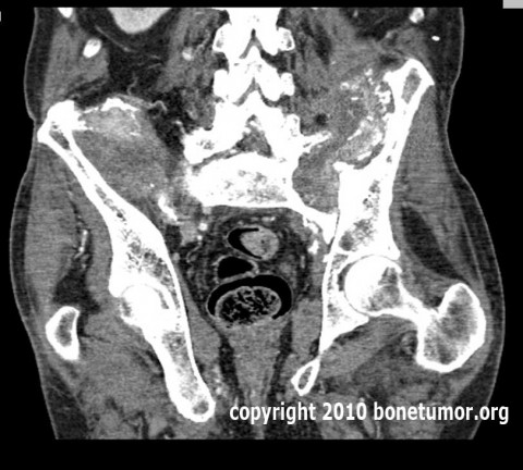
The CT scan shows extensive destructive and reactive changes in the posterior ileum and sacrum bilaterally. There appears to be spinal pelvic discontinuity and subluxation. There appears to have been settling of the spine into the pelvis of several centimeters. The lower extent of the sacral ala on the left side is in contact with the inner table of the pelvis at the level of the left acetabulum, several centimeters below its normal position.
The CT scan shows extensive destructive and reactive changes in the posterior ileum and sacrum bilaterally. There appears to be spinal pelvic discontinuity and subluxation. There appears to have been settling of the spine into the pelvis of several centimeters. The lower extent of the sacral ala on the left side is in contact with the inner table of the pelvis at the level of the left acetabulum, several centimeters below its normal position.
Treatment Options:
The question in this case centers on what can possibly be done to help relieve the patient's pain. Resection of the bone invloved with tumor followed by reconstruction and stabilization of the spine to the pelvis are the possible options.
Resection of the involved areas of bone would be a heroic and risky undertaking, which would not likely result in significant relief of pain. The extensive radiation that has been given virtually guarantees severe wound complications, poor healing, infection, and resulting excess morbidity.
Stabilization of the spine to the pelvis appears to be limited by the extensive damage to the ilia on both sides, thereby severely limiting the bone available for fixation. In addition, the extensive dissection required for implantation of stabilization devices would result in the same risk of severe wound complications due to the high doses of radiation that have been given.
It appears that surgical intervention is not appropriate in this case.
Imagen

Secret Tumor Name
Case ID Number
Image Types
Image modality
Tumor Name
Example Image
yes
Tumor Type
Benign or Malignant
Body region


