Case Identification
Case ID Number
Tumor Type
Body region
Benign or Malignant
Clinical case information
Case presentation
The patient is a very pleasant 86 y/o woman. She says there is absolutely no pain in the thigh She denies any weakness in the right leg. She feels fine, and her appetitie is good, but she has lost 15 lbs in the past few months. Examination of the thigh shows normal mobility of the hip and normal mobility of the knee, and the strength in the two legs is roughly equal.
Radiological findings:
Plain radiographs are shown which are normal.
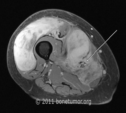
An MRI shows a very extensive right thigh abnormality, that has a somewhat variable appearance. The large lateral and distal portion of the lesion has a relatively homogeneous appearance (see bright areas on some of the MRIs) , the medial and more proximal portions have a less homogeneous appearance (less bright areas on some of the MRIs).
The lesion essentially completely replaces the distal vastus lateralis, extending across the anterior compartment into the vastus medialis, where there were multiple large masses. The lesion also totally encases the femoral artery, and it appears to extend through the medial intramuscular septum into the posterior compartment of the leg, adjacent to but not in contact with the sciatic nerve.
The lesion reaches almost to the knee, and is present in the vastus lateralis just above the knee, and proximally it reaches to the level of the bifurcation of the femoral artery into the profunda femoris and femoral artery. There is also an extension of the lesion into the adductor compartment at this level
Laboratory results:
none that matter
Differential Diagnosis
Sarcoma. Unusual lymphoma?
Further Work Up Needed:
CT scan Chest, abdomen, pelvis, Biopsy
Pathology results:
The pathology images are shown - the first three images are from the large relatively homogeneous distal mass of the tumor, and the second three are from the more heterogeneous proximal area of the tumor. Note the different but related histological features of this tumor.
More homogeneous area: myxomatous - more spindled tumor cells with less pleomorphism and fewer mitoses. More heterogeneous area: epitheloid tumor cells in hyalinized stroma - pleomorphic and numerous mitoses -last three images.
More homogeneous area: myxomatous - more spindled tumor cells with less pleomorphism and fewer mitoses. More heterogeneous area: epitheloid tumor cells in hyalinized stroma - pleomorphic and numerous mitoses -last three images.
Treatment Options:
Is limb salvage an option here?
Special Features of this Case:
This lesion is very unusual in that it presents with very extensive local extent, neurovascular encasement, involvment of the anterior, posterior, adductor compartments and the subcutaneous space of the thigh at presentation, yet other than the size of the mass, there are no symptoms at all. On 7 month follow-up this patient has had 70 Gy radiation, and is doing exceedingly well, still working as a travel agent, Karnofsky performance status 85%. The function, skin condition and appearance, and mobility of the right thigh are virtually perfect. She notes and increasing lump medial to the knee that is 3x3cm and appears to be increasing in size. Additional radiation, although controversial, might be considered.
Image
Secret Tumor Name
Case ID Number
Image Types
Image modality
Tumor Name
Benign or Malignant
Body region


