Case Identification
Case ID Number
Tumor Type
Body region
Benign or Malignant
Clinical case information
Case presentation
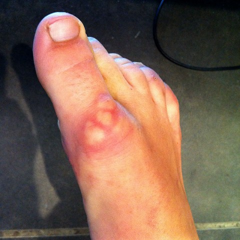
A 22 year old woman had a subcutaneous tumor removed from the top of her big toe 6 months ago. The tumor has already started to come back. A photo from before the surgery is shown.
Radiological findings:
A nonspecific, subcutaneous lesion is seen dorsal to the toe
Laboratory results:
The patient has no findings consistent with rheumatoid arthritis.
Differential Diagnosis
This is a tumor that podiatrists, orthopedic surgeons who treat foot and ankle conditions, and hand surgeons will see relatively frequently . Dermatologists and primary care MD's may also encounter this lesion. Students and practitioners of surgery of the foot should be aware of this lesion and include it in their differential when encountering skin and subcutaneous tumors in the foot.
Further Work Up Needed:
What is the best next step in managing this particular patient?
Pathology results:
The excision shows large areas of geographic necrobiosis with
surrounding palisaded histiocytic inflammation and fibrosis. The areas of
necrobiosis have a somewhat basophilic tint and are shown to contain
degenerated collagen as well as mucin, highlighted on trichrome and Alcian blue
stains respectively. The differential diagnosis for palisaded histiocytic
inflammation in the deep soft tissues primarily includes rheumatoid nodule and
subcutaneous granuloma annulare. Rheumatoid nodules are often seen in older
patients in conjunction with a clinical diagnosis of rheumatoid arthritis. In
addition, mucin deposition within the lesions is unusual, although it has been
reported. Subcutaneous granuloma annulare is more common in children and young
adults, and the areas of necrobiosis show more basophilia and often show mucin
deposition. Unfortunately, there can be histologic overlap between these two
diagnoses. In a young patient in the absence of coexisting rheumatoid
arthritis and given the somewhat basophilic necrobiosis with positive mucin
deposition, the findings are most consistent with subcutaneous granuloma
annulare. However, if the patient has a known history of rheumatoid arthritis
and the lesion is adjacent to an affected joint, these findings are best
interpreted as rheumatoid nodule.
Treatment Options:
This lesion may require biopsy for conclusive diagnosis, but surgical excision is not needed and other treatments are better suited. Surgical treatment is frequently followed by recurrence. See the associated page on this site for complete information on this tumor.
Special Features of this Case:
The patient is a very pleasant 22-year-old woman who presents approximately 6 months after having had surgical treatment of a mass in the left great toe. She shows us a picture of multiple subcutaneous masses, adjacent to each other, on the dorsum of the left great toe, which blanch slightly with flexion of the toe, each may be six or 7 mm and roughly round, apparently projecting from the skin surface by a few millimeters. Overlying skin otherwise normal. These had gradually grown and increased in size over a period of two years.
Image
Secret Tumor Name
Case ID Number
Image Types
Image modality
Tumor Name
Example Image
yes
Benign or Malignant
Body region


