Case Identification
Case ID Number
Tumor Type
Body region
Position within the bone
Periosteal reaction
Benign or Malignant
Clinical case information
Case presentation
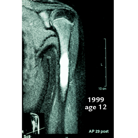
This healthy male was treated for an expansile lesion of the left humeral diaphysis at age 13. At age 25, pain recurred, and xrays show a large new lesion with weakening of the bone.
Radiological findings:
Xrays from the three episodes of treatment are shown, with the most recent xrays shown last. In 1999 at age 12-13 there was an expansile lesion with an incomplete pathological fracture, and bright signal on MRI. Hydroxyapatite cement was used to fill the lesion. In 2006 xrays showed the HA graft material was still present, there was no expansion of the bone or weakening, but an MRI showed persistant mild signal change. In 2011 when the pain recurred, xrays showed a large new expansile lesion in the humerus. The new lesion is proximal to the the HA cement which was placed in 1999. There is no periosteal reaction. The bone is slightly expanded, and the cortex thinned but not violated.
Laboratory results:
The lesion was sampled at surgery, and based on findings of benign tissue, it was curetted and packed with PMMA cement. At surgery, there was slightly turbid, yellow fluid under mild pressure in the lesion, which was send for cytology. Curettings revealed significant amounts of tan cellular material, but also revealed a thin membrane with a shiny, synovial appearance covering portions of the lesion cavity.
Differential Diagnosis
The original pathological material from 1999 was described as "fibroproliferative lesion" but no definite diagnosis was given. Is this an NOF? Is it a recurrent UBC with fibrous proliferation? Or something else?
Further Work Up Needed:
The lesion was sampled, and based on findings of benign tissue, it was curetted and packed with PMMA cement. At surgery, there was slightly turbid, yellow fluid under mild pressure in the lesion, which was send for cytology. Curettings revealed significant amounts of tan cellular material, but also revealed a thin membrane with a shiny, synovial appearance covering portions of the lesion cavity. Pathology is pending
Pathology results:
See images
Special Features of this Case:
This lesion does not fit any classic diagnosis. UBC does not typically occur in the diaphysis, or contain large amounts of tissue, and NOF does not fit the xray appearance, behavior, or the fact that the lesion was fluid filled. FD can undergo cystic change, but usually demonstrates bone formation. None of these lesions typically regrow dramatically as this lesion seems to have done, in a young adult 12 or 13 years after initial treatment.
Image
Case ID Number
Image Types
Image modality
Tumor Name
Example Image
no
Benign or Malignant


