Case Identification
Case ID Number
Tumor Type
Body region
Benign or Malignant
Clinical case information
Case presentation
A 26 year old woman who is 18 weeks pregnant with her 3rd child presents with a very large mass in the right leg. She is otherwise well and her medical history is unremarkable.
Radiological findings:
Xrays are normal, with no calcifications within the mass, and no change in nearby bones. There is mild demineralization of the femur and tibia from disuse. An MRI shows a mass, about 14 x 16 x 28 centimeters in size with mild to moderate heterogeneity, dark T1 anbd brigh T2, centered in the medial gastroc muscle, extending from the proximal popliteal fossa to the mid-distal calf. The neural and vascular structures are not encased, although the popliteal and posterior tibial artery were in contact with the proximal, anterior and medial portion of the mass for approximately 12 or 14 centimeters.
PA Chest radiograph is interpreted as normal
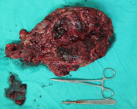
A resection with a marginal, unintentionally contaminated margin was performed. The lesion was very friable, and during the resection, semi-necrotic somewhat myxoid material oozed into the wound and contaminated the field during a portion of the dissection.
PA Chest radiograph is interpreted as normal
A resection with a marginal, unintentionally contaminated margin was performed. The lesion was very friable, and during the resection, semi-necrotic somewhat myxoid material oozed into the wound and contaminated the field during a portion of the dissection.
Laboratory results:
None available
Differential Diagnosis
Sarcoma, synovial versus myxoid liposarcoma or myxoid chondrosarcoma vs partially necrotic other sarcoma.
Pathology results:
pending
Treatment Options:
A resection with a marginal, unintentionally contaminated margin was performed. The lesion was very friable, and during the resection, semi-necrotic somewhat myxoid material oozed into the wound and contaminated the field during a portion of the dissection.
Special Features of this Case:
The patient was treated at the HEODRA hospital in Leon, Nicaragua
Image
Case ID Number
Image Types
Image modality
Tumor Name
Example Image
yes
Tumor Type
Benign or Malignant
Body region


