Case Identification
Case ID Number
Tumor Type
Position within the bone
Benign or Malignant
Clinical case information
Case presentation
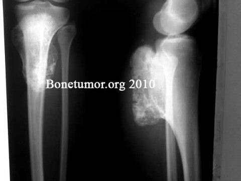
A case sent for consulation. A 22 year old woman presents with a 1-year history of leg pain. No other clinical data are available.
Radiological findings:
A large surface based lesion projects from the posterior and proximal tibia. There is a gradual and mature looking expansion of the posterior cortex of the tibia, which appears to be of long standing. From this cortex projects the mass, very heavily calcified, with a somewhat varigated pattern of calcification, with some ring or round shaped lucent areas.
Laboratory results:
None available
Differential Diagnosis
Exostosis, exostosis with associated low-grade chondrosarcoma, periosteal osteosarcoma, parosteal osteosarcoma.
Further Work Up Needed:
I would recommend a CT scan, and if possible an MRI. The CT scan will show the relationship of the lesion to the bone, and as you may know, the specific appearance of the junction between the lesion and the bone will allow the diagnosis to be made with a high degree of certainty.
Pathology results:
None available
Treatment Options:
Clearly, removal is needed. This is a very difficult place to operate, and a very large tumor, I recommend this patient be operated by a very experienced surgeon or team of surgeons, if that is possible.
Special Features of this Case:
A lesion of exceptionally large size.
Image
Case ID Number
Image Types
Image modality
Tumor Name
Benign or Malignant


