Case Identification
Case ID Number
Tumor Type
Body region
Position within the bone
Periosteal reaction
Benign or Malignant
Clinical case information
Case presentation
The patient is a 62-year-old woman who is seen because of a mass in the right shoulder. She states that her symptoms began approximately two years ago, with stiffness. She had limited motion and was told that she might have a frozen shoulder.
Radiological findings:
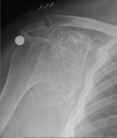
On plain radiographs, there is a large mass in the right scapula. The lesion is a lobular and expansile. It has replaced and expanded the entire glenoid and surrounding bone. The lesion is well-defined, with an extensive ring - like pattern of calcification forming circles, ovals, and rounded shapes within the mass. There is no definite periosteal reaction. The MRI shows a large mass which is dark on T1 and bright on T2. The mass is immediately adjacent to the articular surface of the proximal humerus. The entire glenoid is infiltrated and replaced. The lesion reaches up into the scapular spine. The T2 images show dark interlesional calcifications surrounding bright lobules within the mass. The marrow signal in the proximal humerus is normal.
Laboratory results:
No laboratory examinations are required for diagnosis.
Differential Diagnosis
This large lesion of the scapula bone could be a benign or malignant primary bone tumor. Possibilities include very expansile fibrous lesions, such as fibrous dysplasia, cystic lesions, cartilaginous lesions, and perhaps vascular lesion such as hemangioma. The plain radiographs and MRI findings should be examined for the glue to the diagnosis.
Further Work Up Needed:
The patient will require staging depending on the outcome of the diagnostic process.
Imagen

Secret Tumor Name
Case ID Number
Image Types
Image modality
Tumor Name
Tumor Type
Benign or Malignant
Body region
Bone name
Location in the bone
periosteal reaction
position within the bone
Tumor behavior
Tumor density


