Case Identification
Case ID Number
Tumor Type
Body region
Benign or Malignant
Clinical case information
Case presentation
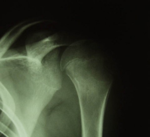
An 11 year old boy had pain in the left shoulder. A left proximal humeral lesion was noted on plain radiographs and CT images, which are shown.
Radiological findings:
A biopsy followed by curettage and packing of the lesion with methylmethacrylate cement was carried out, also shown in the images. The microscopic appearance of the tissue is shown. One year later he again complained of pain, and a repeat CT was performed. The new CT images are shown.
Pathology results:
The microscopic appearance of the tissue is shown.
Treatment Options:
1) What is the diagnosis?
2) What is the cause of the pain that began one year post-operative?
3) What should be done now?
2) What is the cause of the pain that began one year post-operative?
3) What should be done now?
Imagen

Secret Tumor Name
Case ID Number
Image Types
Image modality
Tumor Name
Benign or Malignant


