Case Identification
Case ID Number
Tumor Type
Benign or Malignant
Clinical case information
Case presentation
The patient is a 10-year-old girl who has a history of pain in the right wrist for approximately six weeks. She also notes pain in the left knee. There is no definite history of injury.
Radiological findings:
Examination of the right upper extremity reveals a little bit of warmth and a little bit of thickening or swelling of the entire wrist. There is no definite mass. There is no lymphadenopathy in the elbow nor in the axilla.
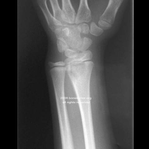
Radiographs of the right wrist and on the left knee are made. There is a lytic lesion in the distal radius and adjacent to the distal radial ulnar joint. It reaches up to the growth plate. There is a subtle periosteal reaction. The lesion has very high signal intensity on T2 MRI images. Images of thge left knee are normal.
Radiographs of the right wrist and on the left knee are made. There is a lytic lesion in the distal radius and adjacent to the distal radial ulnar joint. It reaches up to the growth plate. There is a subtle periosteal reaction. The lesion has very high signal intensity on T2 MRI images. Images of thge left knee are normal.
Laboratory results:
The laboratory examination shows a mildly elevated white count, and elevation of the erythrocyte sedimentation rate and the C-reactive protein. Bacterial cultures of the bones and blood are negative.
Image

Case ID Number
Image Types
Image modality
Tumor Name
Tumor Type
Benign or Malignant
Body region
Bone name
Location in the bone
periosteal reaction
position within the bone
Tumor behavior
Tumor density


