Case Identification
Case ID Number
Tumor Type
Body region
Benign or Malignant
Clinical case information
Case presentation
The patient is 74, with pain and small area of swelling on the left foot for 2 months. The swelling has increased and a definite mass is present. The patient has prostate cancer treated years ago. He quit smoking 40 years ago.
Radiological findings:
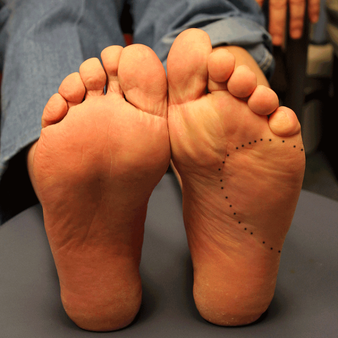
Physical examination shows a definite visible and palpable mass, that is present on the dorsal extensor surface and in a more subtle way on the plantar surface of the lateral portion of the left foot as well (see photos). The mass is slightly tender, slightly warm, no evidence of infection. Very slight redness is seen. The patient prefers roomy slippers because his shoes are too painful (see photos). He is able to ambulate but he has pain that has required ongoing medication with Motrin or Tylenol for the past couple of weeks. The pain is present both day and night.
The xrays show some subtle findings (see photos). There is slight loss of the cortex on the lateral side of the fourth and there is some change on the cortex on the medial side of the fifth. The mass itself does not have calcifications.
The MRI shows a quite large mass, measuring five or 6 cm x 5 or 6 cm in the dorsal aspect of the foot, and about 7 x 7 or 8 x 8 cm in the plantar aspect of the foot, somewhat dumbbell shaped in overall appearance, extending through the fourth interspace (see photos). It is bright on T2, and dark on T1. Based on the MRI, it looks like this mass started as a soft tissue mass between the fourth and fifth metatarsals and gradually spread into the dorsal soft tissues, the plantar soft tissues, and gradually began to erode the nearby cortex of the fourth and fifth metatarsals.
Chest x-ray shows three nodules, in the right upper lobe 12.7 mm, 17 mm in the left lower lobe, 26 mm near the lingula.
Laboratory results:
The patient's prostate specific antigen is normal. Complete blood count is normal. Kidney function tests are normal. Liver enzymes normal. Chest x-ray shows three nodules, in the right upper lobe 12.7 mm, 17 mm in the left lower lobe, 26 mm near the lingula.
Differential Diagnosis
This lesion appears to be a soft tissue mass. There are nodules in the lung which could represent metastasis. It is less likely that the nodules in the lung are a primary and the lesion the foot is a secondary or metastatic lesion. The history of prostate cancer is significant, but the PSA is low ruling out prostate cancer. Thyroid function is normal, effectively ruling out thyroid metastasis to the bone.
Further Work Up Needed:
Biopsy is necessary.
Pathology results:
pending
Image

Secret Tumor Name
Image Types
Image modality
Tumor Name
Example Image
yes
Benign or Malignant
Body region


