Evaluation of the Risk of Pathologic Fractures Secondary to Metastatic Bone Disease
Pathologic fractures create a serious morbidity in patients with metastatic bone disease. Orthopedic surgeons who treat patients with metastatic skeletal lesions should focus on proactive treatments designed to prevent pathologic fractures before they occur. Prevention of pathologic fractures result in better patient outcome, lower cost, and less difficult operative procedures. For this reason, it is critical to identify both patients and skeletal lesions that are at increased risk of pathologic fracture. The goal of this review is to establish a systematic screening tool and treatment algorithm that orthopedic surgeons can easily apply to their patients in order to optimize the management of metastatic skeletal disease.
Unlike fractures of normal bone, pathologic fractures occur during normal activity or minor trauma due to weakening of the bone by disease. Conditions associated with pathologic fractures include underlying metabolic disorders, primary benign tumors, and primary and metastatic malignant tumors (q) . The most common condition associated with pathologic fractures is osteoporosis(q). This review will focus on the evaluation of fractures that occur secondary to bone destruction by metastatic cancer. Prevention of pathologic fractures is superior to treatment after the fact. Some of the advantages that have been cited include shorter hospital stays(R,A); easier rehabilitation and nursing with more rapid restoration of function (U,V,K,R); easier radiotherapy treatment (R,A.); more immediate pain relief (U,V,K,R,A); and faster and less complicated surgery (R,A).
In order to determine which patients require prophylactic fixation to prevent pathologic fracture, it is necessary to perform an accurate and reliable risk evaluation. Many different characteristics have been proposed as important criteria for determining risk of fracture. These include type of cancer; type of treatment; size of the lesion; location of the lesion; whether the lesion is lytic or blastic; and symptoms due to the lesion. In addition, some have proposed a detailed biomechanical analysis based on finite element modeling. the use of biomechanics to predict fracture. This article will critically review the literature and provide guidelines for estimating fracture risk that are useful for orthopedic surgeons.
PATIENT FACTORS
Cancer Diagnosis The patient's underlying cancer diagnosis is an important component of their pathologic risk profile (Table 1). Breast cancer is the most important source of bone metastasis, and it is responsible for the majority of the skeletal metastases that require orthopedic consultation (M). The risk of pathologic fracture increases with the duration of metastatic disease. Because breast carcinoma has a relatively long survival, these patients are more likely to sustain a pathological fracture. Based on the author's experience, breast cancer metastases that are purely lytic are more likely to fracture than those that are blastic or mixed lytic and blastic. However, blastic lesions in high risk areas such as the proximal femur have a high rate of fracture.
Prostate cancer, combined with breast cancer, contributes to 80% of all skeletal metastasis (O). Prostate cancer normally forms blastic metastases which are less susceptible to fracture, but blastic lesions have been shown to decrease the longitudinal stiffness of bone (?). In addition, some of the treatments that are commonly given for prostate cancer increase the likelihood of pathologic fracture. These include LHRH agonists, orchiectomy, and radiation. In one study, patients receiving LHRH agnosists had a 9% incidence of fracture, a rate significantly higher than similar patients not receiving LHRH agonists (Cancer 1997, February 1st, Volume 79 (3), Pg 545). Patients with prostate cancer who have had radiation to bony areas, or who have low bone density due to hormone modification therapies should be considered at increased risk for fracture.
Lung cancer has a relatively aggressive course and a short survival after bone metastasis. Thus fewer patients survive long enough to develop pathologic fracture. Metastases are typically lytic and have a correspondingly higher risk of fracture. A small proportion of lung cancer metastasis can occur in bones below the elbow and the knee (acrometastasis). These lesions are frequently painful and require radiation or surgical treatment due to the pain rather than for risk of fracture as the risk of functionally disabling fracture through an acrometastasis is low.
Bone metastasis is diagnosed in 4% - 13% of patients with thyroid cancer (Marcocci et al, Surgery 106:960-966, 1989 and McCormack, Cancer 19:181-184 1965.) The lesions are frequently lytic and their fracture risk depends on their location. Because patients with thyroid cancer may have prolonged survival they are also at increased overall risk of pathological fracture. Approximately 25-50% of renal cell carcinomas metastasize to bone (r,s).
Renal cell metastases to bone can be unusually expansile and destructive, which creates an increased risk of pathologic fracture. Orthopedic surgeons treating metastatic cancers should note that certain selected patients with renal cell metastases may be candidates for aggressive surgical resection for cure (?). Table 1: Origin and Rates of Metastasis to Skeleton Irradiation of Lesion
Irradiation of metastatic bone lesions also appears to increase the risk of pathologic fracture (C, N, 1, G, R, K,A,Z). Keene et al found that 18% of patients who underwent irradiation for metastatic breast carcinoma developed fractures. Other authors have reported much higher incidences ranging from 26%-41% (1,N,Z). Harrington (G) theorizes that radiotherapy increases the risk for fracture because it causes temporary softening of the bone at the tumor site. Radiation may lead to increased fracture risk due failure of reossification after treatment. Beals and Snell reported that only 4% of lesions reossified after treatment (A). However, other authors have found a 65%-85% incidence of reossification under similar circumstances, assuming a fracture has not occurred (4, Q).
Pain Pain is an important but controversial criteria for evaluation of pathologic fractures. In metastatic disease, pain may arise from enlargement of the tumor, perilesional edema, increased intraosseous pressure, or weakness from bone loss (1,X). The direct pressure exerted by the tumor on the bone has been shown to stimulate the release of various pain mediators including porstaglandins, bradykinins, and histamine (o). In addition, tumor invasion of bone can lead to activation of mechanoreceptor and nociceptors which leads to the development of pain (o). The controversy lies in whether or not pain can be used as a sign of impending fracture. Fidler (B) stated that pain could not be considered a reliable sign of an impending fracture because only half of the patients in his study complained of pain. Keene et al (C) found that most patients with metastatic bone cancer did develop bone pain, but only 11% of them actually had fractures; therefore, he concluded that pain was not an accurate indication of impending fracture.
Many authors (A,D,E,F,G, R,1) feel that pain is an important indication for prophylactic fixation. Some have singled out persistent pain despite radiation (D,E,G) as a criterion for fixation while others state that pain caused only by lytic lesions should undergo prophylactic fixation (F). In some series, patients without pain had a low risk of fracture ( R) and patients with functional pain had a high risk of fracture approaching 100% (R, 1). These finding suggest that pain may be a valuable sign of decreased mechanical strength of bone and increased fracture risk (1).
LESION FACTORS
Relationship of Lesion Size to Fracture Risk Beals and Snell (A) published highly influential works in 1956 and 1961. Their work dealt only with patients with breast cancer and only with lesions in the femur. Of the 19 fractures that occurred in their first series, they found that 58% of these fractures were predictable using the following criteria: Presence of a metastatic lesion 2.5 cm in size or larger involving the femoral cortex or presence of a defect of the same size in any location that caused pain to the patient. These criteria were used in a second series and patients at risk for fracture underwent prophylactic fixation of the diseased bone. This treatment reduced the incidence of fracture from 32% to 9%.
Parrish and Murray (J,E) used these criteria as indications for prophylactic fixation when performing their studies on treatments of pathologic fractures. They found that using these criteria led to decreased fractures and improved the quality of life of their patients. In 1973, Fidler(B) retrospectively studied 19 patients with pathologic fractures of the femur. He found that 100% of patients with greater than 50% cortical involvement developed a fracture. Based on this data, he recommended that patients with involvement of over half of the cortex should undergo surgery to stabilize the bone. In 1981, Fidler (D) published another retrospective study of 66 patients with 100 metastases in the long bones. His results corroborated his previously reported indications for prophylactic fixation. He found that when greater than 75% of the cortex is destroyed the incidence of fracture is 80%. When less than 50% of the cortex is involved, the incidence is only 2.3%.
Zickel and Mourandian(P) studied 34 patients with lesions in the proximal femur. They concluded that involvement of even small parts of the cortex in the subtrochanteric region places the femur at high risk for fracture and warrants prophylactic fixation. According to their results, size of the lesion did not correlate with risk of fracture. Keene et al supported this conclusion as he found that all the measureable lesions that fractured had a similar extent of cortical involvement as those that did not fracture ( C).In 1982, Harrington (4) recommended intramedullary nail fixation when there is greater than 70% destruction of the cortex (G).
Table 2: Summary of Publications Studying Effect of Lesion Size on Fracture Risk Author(yr. Published) Criteria evaluated Comments Study Design Beals and Snell (1956, 1961) Size of lesion -Lesion 2.5 cm or larger involving femoral cortex or defect of same size in any location that caused pain predicted occurrence of fracture 1956-retrospective, 19 pts 1961-prospective, clinical trial, Parrish and Murray (1970) Lesion ³ 2.5 cm -This criteria predicted fracture occurrence Retrospective, 96 pts Fidler (1973) Degree of cortical involvement -Prophylactic surgery for those with involvement of >50% cortex Retrospective, 19 patients Zickel and Mourandian (1976)
Location of Lesion -Any involvement of cortex in subtrochanteric region of femur increased fracture risk. -Size did not correlate with fracture risk Fidler (1981) Degree of cortical involvement -Confirmed recommendation for prophylactic surgery for those with involvement of >50% cortex Retrospective, 66 pts, 100 metastases in long bones Harrington (1982) Degree of cortical involvement -Prophylactic fixation when have >70% cortical destruction Mirels (1989) Site of lesion Pain associated with lesion Type of lesion (lytic vs blastic) Size of lesion -Use combination of site, pain, type, size of lesion to determine indication for prophylactic fixation -see Table 3 for scoring system Retrospective, 78 bone lesions
In 1989, Mirels (1) developed a scoring system to quantify the risk of pathologic fracture based on a retrospective study of 78 irradiated metastatic bone lesions. Unlike all the previous studies, Mirels combined four different features of bone lesions in an attempt to create a more reliable risk assessment (See Table 3).
Table 3. Mirels' Scoring System Score variable 1 2 3 Site Upper limb Lower limb Peritrochanter Pain Mild Moderate Severe Lesion Blastic Mixed Lytic Size <1/3 1/3-2/3 >2/3Prophylactic Fixation indicated for score of ³ 9
His system assigned points to the following four variables: the location of the lesion (upper limb, lower limb, peritrochanter); the degree of pain caused by the lesion (mild, moderate, severe); the type of lesion (lytic, blastic, mixed); and the size of the lesion (<1/3, 1/3-2/3, >1/3). Adding the points from each category determines the score. His data indicated that a score of less than or equal to 7 out of 12 is indicative of a lesion not at risk for fracture. A score of 8 out of 12 is associated with a 15% risk for fracture. The risk of fracture is 33% in patients with a score of 9. Mirels concluded that a score of 9 or greater should be used as an indication for prophylactic fixation. Mirels found that the combined score was a more accurate predictor of fracture than any of the four factors used separately. Pain and lesion size were more accurate predictors than type of lesion or site of lesion. Confirming Fidler's conclusions, Mirels found that the rate of fracture was only 5% when the lesion was less than two thirds of the diameter of the bone, but increased to 81% when the lesion was greater than two thirds of the diameter of the bone.
Accuracy of Lesion Size Measurement Several authors have demonstrated the limitations of using size-based criteria alone. In 1986, Keene et al (C) performed a retrospective study of 203 female patients with 516 metastatic breast cancer lesions that were located in the proximal femur. They showed that 57% of the metastases could not be accurately measured from plain radiograph because they lacked a clear border between the lesion and normal bone. Size-based criteria may not be applicable to bony lesions where the cortex cannot be effectively measured, such as the spine, ribs, and pelvis (N).
Hipp et al also stated that characteristics of both metastatic bone lesions and physician observers lead to a very high degree of error and variability in the measurement of lesions (5). Two physicians reading the same radiograph and applying the same criteria might come to very different conclusions about the need for prophylactic fixation, with potentially disastrous consequences for the patient. In fact, Hipp et al found that experienced orthopedic oncology surgeons could not consistently predict strength reductions or load bearing capacity from radiographs or CT films (5). Therefore, another method needs to be found to determine factor of risk that would be easier for orthopedic surgeons to use.
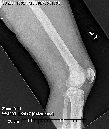
Relationship of Lesion Location to Fracture Risk Of the long bones in the peripheral skeleton, the femur is the most common site for metastases followed by the humerus (R,U). According to Knutson et al, 88.4% of all long bone metastasis secondary to breast cancer involved the femur (R). Within the long bones, the proximal part is most likely to be affected, especially the peritrochanteric region of the femur (1, U,d). While metastases to the long bones account for less than 20% of all fractures (O), Harrington found that over half of these pathologic long bone fractures occurred in the proximal femur (G). Some authors have suggested that the femur is more likely to sustain a pathologic fracture than other long bones (A,R,T,U,V). Dijkstra et al states that 25% of all long bone metastases fracture, but the proximal femur has an incidence of 40-60% (U).
However, Fidler (D) found no difference between the rate of fracture in upper limb lesions versus lower limb lesions. Mirels also found that the peritrochanteric region, while the most common area of the femur to develop metastases, is not any more likely to fracture than other sites (1). Taking into consideration the conflicting information in the literature, it is the authors opinion that the orthopedic surgeon treating metastatic skeletal lesions should have a relatively low threshold for prophylactic fixation of proximal femur lesions. Any lesion between the lesser trochanter and the femoral head causing functional pain or larger than 2.5 cm should be fixed prophylactically. Pathologic fractures in this location produce serious morbidity. The operative procedures applicable to the proximal femur are familiar to all practicing orthopedic surgeons. The benefit of prophylactic fixation significantly outweigh the risks of surgery.
Lytic vs. Blastic Lesions While bone formation and destruction occur simulataneously in most metastatic cancers, usually one predominates over the other. Mirels (B) and others (4,f,P,N) have found that lytic lesions did have a higher risk for fracture. Mirels found in his study that none of the blastic lesions fractured, but 32% of the mixed lesions and 48% of the lytic lesions did. He theorized that lytic lesions were a result of a more advanced process of bone resorption . On the other hand, Hipp et al (g) found that even though blastic lesions do increase bone density, they do not change bone strength and they decrease the stiffness. Lytic lesions decrease both strength and stiffness of the bone.
Biomechanical Modeling of Fracture Risk Biochemical testing and computer modeling have contributed to the understanding of fracture risk. Hipp et al (5) discusses in vitro experimentation as an alternative to using clinical and radiographical data to predict pathologic fractures. Studies have shown that even small cortical defects can significantly reduce the strength of the bone (h,j,m). Brooks et al found that drill holes as small as 2.8 mm or 3.6 mm in the femoral mid-shaft significantly weakened the bone because of increases in local stresses by the defect (h). Hipp found that a hole that reduced the cross-sectional area of the bone by less than 40% reduced the torsional strength of the bone by 70%. These results suggest that the 50% loss originally stated by Fidler (B) as the cutoff for prophylactic fixation may be an underestimation. Hipp et al (l) also found that the location and shape of endosteal defects affected the degree of strength reduction in the bone which would therefore affect the risk of fracture. If a defect causing a 50% loss in cross sectional area is in the center of the femoral diaphysis, the strength of the bone is reduced by 60%. However, if an identical defect is located such that the thinnest wall was at the point of maximal bending stress, the strength reduction was greater than 90%.
In bones subjected to bending, it is the location of the defect that is important in determining the amount of strength reduction. The length of the defect along the long axis of the bone has a large effect on torsional strength. A long defect with the same decrease in cross sectional area as a small defect will cause a greater reduction in strength than the smaller defect. The length of the defect does not significantly affect bone strength if bones are subjected to bending (5,l). Studies need to be conducted that study the effect of combinations of torsion and bending to determine how they affect the strength of the bone. Biomechanics and computer models promise to improve the accuracy of fracture risk prediction, but these methods are not yet available for everyday use.
Summary In summary, an orthopedic surgeon calculating risk of pathologic fracture is likely to focus most of his or her attention on the appearance of lesion of plain radiographs. The authors recommend that the size of the lesion be considered in the context of the other factors that are mentioned by Mirels et al (1). When the boundaries, or dimensions, of a lesion are uncertain, the threshold for orthopedic stabilization should be lowered.
In certain locations such as the femoral neck, the peritrochanteric region of the femur, and the junction between the humeral head and the humeral metaphysis, the risk and disability from pathologic fracture are so great that orthopedic stabilization should be used in virtually all cases. Only very small, well-delineated lesions in these high-risk locations should be treated non-operatively. If the surgeon chooses non-operative care for a small lesion in a high risk location, careful follow-up is required since the lesions may progress to fracture before the treatment is completed.
RECOMMENDATIONS
There is no doubt about the importance of determining which cancer patients with metastatic bone disease have had enough damage to their bones to increase their risk of developing a fracture. Prophylactic fixation of these patients clearly decreases morbidity in patients compared to fixation of completed fractures. The difficulty lies in determining a good set of criteria that allows surgeons to accurately determine the patient population requiring prophylactic fixation. Many different criteria have been suggested including the size of the lesion; the type of cancer that metastasized to bone; the location of the metastatic lesion; pain due to the lesion; whether the lesion is lytic or blastic; irradiation of the lesion; and the use of biomechanics to predict fracture.
However researchers have disagreed on which are the important features to use for diagnosis of imminent fractures as in most cases there has been evidence published that supports and that refutes the use of each of the features for diagnosis. When deciding which criteria to use, it is important to consider both the accuracy with which it predicts an increased risk for fracture and the convenience with which it can be measured. The biomechanical factors that Hipp proposes to use seem as though they would be good predictors of fracture, but Hipp did not provide an easy way to collect the data required making them less useful as a diagnostic tool. While there is evidence that the size of the lesion could be a good predictor for fracture, the difficulty in accurately determining the size radiographically makes it less useful diagnostically.
The other variables discussed are much easier to determine in patients and therefore would seem more useful diagnostically. However, based on the research it seems that one criteria alone is not accurate enough to predict and increased risk of fracture. Rather, having the requirement for fixation involve the presence of several of these criteria would be better because while each criteria individually is subject to error by the physician, probability indicates that the chance of having false negatives and false positives would decrease with the more factors involved. Therefore, this writer feels that, while not ideal, the best diagnostic system to use is the scoring system proposed by Mirels who requires the analysis of 4 criteria in determining risk of fracture. In addition, his research indicates specific scores over which patients should have prophylactic surgery.
Therefore, surgeons would be able to relatively easily determine which of the criteria their patient has, assign them a score, and decide whether to perform surgery based on the score and clinical suspicion. While this system seems to be the best for now, it is likely that with improvements in imaging studies and further research, new criteria and modifications of old criteria will soon be proposed. . Metastatic bone disease is the most common malignant bone lesion seen in adults. Bone is the third most common site for metastases after the lung and the liver (H,I). 7-27% of all cancer patients each year are likely to have a metastatic bone defect (H). The incidence of pathologic fractures in patients with malignant disease is 1-2% and 25% of all metastases to long bones progress to fractures (U).