Case Identification
Case ID Number
Tumor Type
Body region
Position within the bone
Benign or Malignant
Clinical case information
Case presentation
The patient is a very pleasant 45-year-old banker who has noticed a mass in the right foot for approximately 6 months.
Radiological findings:
On oncologic examination, the overall status, and regional status of the lesion is assessed. There is a palpable lymph node in the right inguinal ligament area, and no lymphadenopathy in the right popliteal fossa. No central lymphadenopathy is noted. There is no café au lait spot, or other unusual skin lesion. The patient does not appear chronically ill or cachectic.
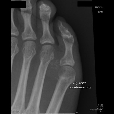
In the foot, there is a firm, somewhat tender, deep, fairly sizable mass that surrounds the neck of the distal portion of the fifth metatarsal, spreading into the interspace between the fourth and the fifth metatarsal. The dorsal side of the foot is slightly swollen. There is slight lifting up of the lateral border of the foot because of the bulk of the mass. The overlying skin circulation as well as the neurologic findings are normal. There is no definite Tinel's sign but the mass is quite tender and percussing the mass does cause pain.
The plain radiographs of the foot show a few foci of calcification within the mass. Click on the images to see larger views.
MRI shows a multiloculated mass surrounding the fifth metatarsal shaft near the distal end. It has grown between the fourth and fifth up into the dorsal portion of the foot and is apparent between the fourth and fifth toes and in the tendinous interspace of the fifth. Click on the images to see larger views.
CT scan of the chest, abdomen and pelvis shows bilateral inquinal adenopathy, with multiple small bilateral nodes, but no other finding of concern.
Imagen

Case ID Number
Image Types
Image modality
Tumor Name
Tumor Type
Benign or Malignant
Body region
Bone name
Location in the bone
position within the bone
Tumor behavior
Tumor density


