Case Identification
Case ID Number
Tumor Type
Body region
Position within the bone
Periosteal reaction
Benign or Malignant
Clinical case information
Case presentation
Patient has a mass he thinks might be a varicose vein on the anterior surface of his left leg for several years.
Radiological findings:
An x-ray shows that there is a lesion in the mid tibia which corresponds with the soft tissue abnormality noted. It appears that there is a series of openings in the tibial cortex at the location of the lesion, the largest of which is less than a centimeter in length and perhaps 0.5-cm in width. There are couple of smaller nearby openings as well. Overall the size of the lesion represents a little bit more than half the diameter of the shaft of the bone. There is no periosteal reaction. There is no matrix mineralization or other visible abnormality on x-ray.
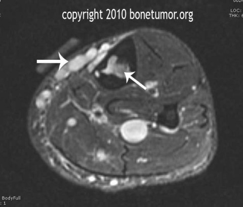
The MRI shows what appears to be a vascular anomaly. The lesion can be clearly seen crossing the tibial cortex, line both anterior and the tibial cortex perhaps underneath the periosteum, transgressing the cortex, and then lying within the intramedullary space. It has the globular appearance of a possible vein, and it connects to an elongated vascular appearing structure that exits proximally through a nutrient foramen in the posterior tibial cortex and joins the vascular elements in the soft tissues behind the tibia. The size of the vessel behind the tibia is slightly enlarged as well.
The MRI shows what appears to be a vascular anomaly. The lesion can be clearly seen crossing the tibial cortex, line both anterior and the tibial cortex perhaps underneath the periosteum, transgressing the cortex, and then lying within the intramedullary space. It has the globular appearance of a possible vein, and it connects to an elongated vascular appearing structure that exits proximally through a nutrient foramen in the posterior tibial cortex and joins the vascular elements in the soft tissues behind the tibia. The size of the vessel behind the tibia is slightly enlarged as well.
Differential Diagnosis
Vascular tumor vs vascular anomaly
Special Features of this Case:
The MRI appearance of this lesion appears to correspond exactly with the entity described as interosseous venous drainage anomaly. This has been described in the literature on a few occasions and a handful of cases have been reported.
Reference:
Intraosseous venous drainage anomaly of the tibia treated with imaging-guided sclerotherapy.
Peh WC, Wong JW, Tso WK, Chien EP.
Br J Radiol. 2000 Jan;73(865):80-2.
Intraosseous venous drainage anomaly in patients with pretibial varices: imaging findings.
Boutin RD, Sartoris DJ, Rose SC, Plecha EJ, Bundens WP, Haghighi P, Harter LP, Resnick D.
Radiology. 1997 Mar;202(3):751-7.
Image
Secret Tumor Name
Case ID Number
Image Types
Image modality
Tumor Name
Tumor Type
Benign or Malignant
Body region


