Case Identification
Case ID Number
Tumor Type
Body region
Position within the bone
Periosteal reaction
Benign or Malignant
Clinical case information
Case presentation
The patient has a history of pain in the hip that dates back about two months. The pain became gradually worse and he had some small episodes of exacerbation. Recently he began using crutches. Then, he had another episode of a slight twisting injury with dramatic increase in the pain and was found to have a fracture.
Radiological findings:
He is 26 1/2 years old. He is otherwise generally healthy. The patient denies any fevers chills weight loss anorexia or other systemic symptoms.
Abdominal examination shows no mass, and there is no inguinal lymphadenopathy. There is shortening of the right leg versus left of about 1 1/2 inches. There is pain with motion. There is no generalized swelling.
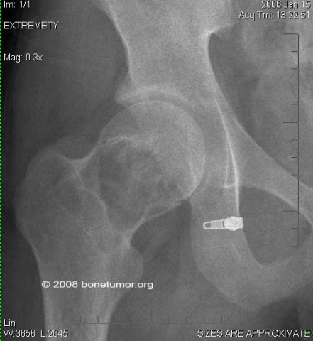
Radiographs show a lytic destructive lesion in the right proximal femur which has a relatively sharp well marginated border and there is no definitive soft tissue mass.
MRI shows a well defined mass in the femoral neck and head with bright signal on T2 and dark signal on T1, with very little reacive change and no apparent soft tissue mass.
A workup shows no abnormaliy in the chest, abdomen, or pelvis other than what is shown. Bone scan shows this lesion appears to be solitary.
Abdominal examination shows no mass, and there is no inguinal lymphadenopathy. There is shortening of the right leg versus left of about 1 1/2 inches. There is pain with motion. There is no generalized swelling.
Radiographs show a lytic destructive lesion in the right proximal femur which has a relatively sharp well marginated border and there is no definitive soft tissue mass.
MRI shows a well defined mass in the femoral neck and head with bright signal on T2 and dark signal on T1, with very little reacive change and no apparent soft tissue mass.
A workup shows no abnormaliy in the chest, abdomen, or pelvis other than what is shown. Bone scan shows this lesion appears to be solitary.
Laboratory results:
no significant findings
Image
Secret Tumor Name
Case ID Number
Image Types
Image modality
Tumor Name
Benign or Malignant
Body region
Location in the bone
periosteal reaction
position within the bone
Tumor behavior
Tumor density


