Case Identification
Case ID Number
Tumor Type
Body region
Position within the bone
Periosteal reaction
Benign or Malignant
Clinical case information
Case presentation
This very pleasant 55-year-old woman has a lump in the left breast and pain in the left hip. She found the breast mass 1 year ago, and it has gotten larger. She first noticed gradually increasing pain in the left groin 8 or 9 months ago. Now, the patient cannot walk without crutches. She has extreme pain with certain activities, and is taking large doses of narcotic pain medicines.
Radiological findings:
On past medical history, the patient had a benign lump removed from the right breast in the 1980s. She does not smoke.
On oncologic examination, the patient does not appear chronically ill or cachectic. There is a mass, which is firm and nontender, in the left breast laterally which measures about 4 x 4 cm.
On musculoskeletal examination, the right hip has a very restricted range of motion. Although the patient to walk, she is very great difficulty placing any weight on the right leg. She can sit, but she cannot lay back without bending the knee up because extending the right hip is very painful. From the flexed position, the hip can be gently abducted without too much pain. No masses palpable along the proximal femur or in the distal femur near the knee.
Plain films show a lytic, permeative, destructive lesion that seemed to be centered in the cortex of the right femur just below the lesser trochanter. Part of the cortex is damaged. There is poorly defined lytic process extending into the intertrochanteric area and down the proximal portion of the femur for 8 or 10 cm.
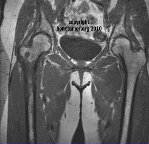
An MRI shows a permeative destructive lesion in the same location proximal right femur, with no soft tissue mass. The bone scan shows multiple abnormal areas of tracer uptake, the proximal femur as a large area of abnormal uptake, there are at least two lesions in the ribs, there are multiple scattered lesions in the right distal femur, and there is one lesion in the pelvis.
On oncologic examination, the patient does not appear chronically ill or cachectic. There is a mass, which is firm and nontender, in the left breast laterally which measures about 4 x 4 cm.
On musculoskeletal examination, the right hip has a very restricted range of motion. Although the patient to walk, she is very great difficulty placing any weight on the right leg. She can sit, but she cannot lay back without bending the knee up because extending the right hip is very painful. From the flexed position, the hip can be gently abducted without too much pain. No masses palpable along the proximal femur or in the distal femur near the knee.
Plain films show a lytic, permeative, destructive lesion that seemed to be centered in the cortex of the right femur just below the lesser trochanter. Part of the cortex is damaged. There is poorly defined lytic process extending into the intertrochanteric area and down the proximal portion of the femur for 8 or 10 cm.
An MRI shows a permeative destructive lesion in the same location proximal right femur, with no soft tissue mass. The bone scan shows multiple abnormal areas of tracer uptake, the proximal femur as a large area of abnormal uptake, there are at least two lesions in the ribs, there are multiple scattered lesions in the right distal femur, and there is one lesion in the pelvis.
Special Features of this Case:
The risk of pathological fracture can be calculated according to a specific method, called the Mirel's score. What is the score and what is the risk of fracture?
Image
Secret Tumor Name
Case ID Number
Image Types
Image modality
Tumor Name
Tumor Type
Benign or Malignant
Body region


