Case Identification
Case ID Number
Tumor Type
Body region
Position within the bone
Benign or Malignant
Clinical case information
Case presentation
This woman had a biopsy last year by a visiting medical team, but no one informed her of the diagnosis. She says the mass is larger. Moderate pain. No inflammation. Well healed scar.
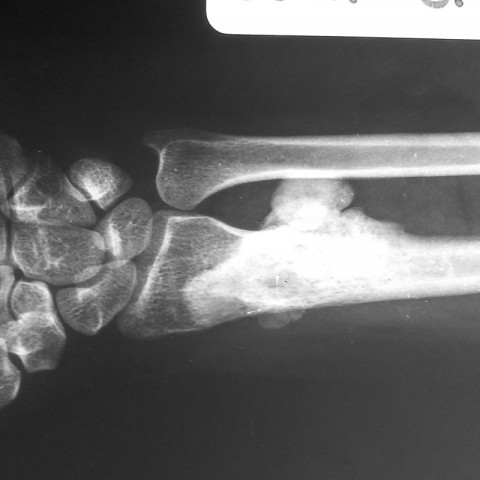
Radiological findings:
A dense, sclerotic, well defined bony mass is seen in the distal radius which extends throughout the metaphysis and distal diaphysis. There is a large extraosseous mass which is densely calcified and lobular that projects to the dorsal and ulnar side of the bone. There is no periosteal reaction around the mass.
Differential Diagnosis
Bone forming tumor. Not a surface lesion (OCE). Osteoblastoma, low grade osteosarcoma (parosteal osteosarcoma).
Image
Case ID Number
Image Types
Image modality
Tumor Name
Example Image
yes
Benign or Malignant
Body region
Bone name
Location in the bone
periosteal reaction
position within the bone
Tumor behavior
Tumor density


