Case Identification
Case ID Number
Tumor Type
Body region
Position within the bone
Benign or Malignant
Clinical case information
Case presentation
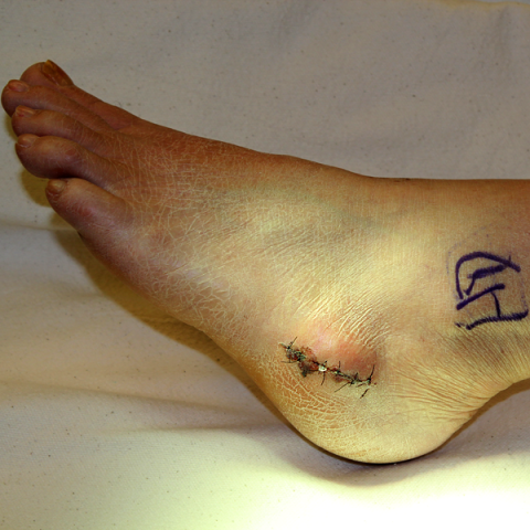
The patient reports an enlarging mass in the left foot for 2 years. Apparently it was small 2 years ago and now is quite substantial. An open biopsy was done, and resection is now planned.
Radiological findings:
There is a bony and cartilagenous mass on the plantar lateral surface of the calcaneus. Once the surrounding tissues are freed, the mass measures more than 8 cm. from base to the most distal point (arrows). At the base is a stalk at its base where the cortex of the bone and the lesion are confluent. The normal trabecular bone of the medullary portion of the calcaneus continues into the lesion. There is thick cap with several large lobules of cartiagenous tissue at the furthest extent definitely more than 2 cms thick (see images). Osteotomy is carried out through normal bone within the body of the calcaneus, resecting the entire stalk of the lesion and its cap en bloc.
Differential Diagnosis
chondrosarcoma vs osteochondroma
Special Features of this Case:
Lingering concerns about the nature of this lesion necessitated an ample incision for complete exposure and an aggressive resection.However, the peroneal tendons (arrows), a portion of the plantar fascia (arrow), and the tuberosity of the calcaneus were spared, and primary closure was achieved without difficulty.
Image

Secret Tumor Name
Case ID Number
Image Types
Image modality
Tumor Name
Example Image
yes
Tumor Type
Benign or Malignant
Body region
position within the bone


